How a calcium assay actually helps us understand what is going on inside living cells
The hidden language of calcium in our cells
When we think about calcium, most of us immediately picture strong bones and healthy teeth. While that is certainly true, calcium plays an even more dynamic role deep within our bodies that most people never see. It acts as a universal messenger, a sort of biological ‘switch’ that tells cells when to move, when to grow, and even when to die. Because this ion is so central to life, scientists have developed sophisticated ways to track it. This is where the calcium assay comes into play, serving as a vital window into the secret life of cells.
At its core, a calcium assay is a laboratory method used to measure the concentration of calcium ions within a sample, usually inside living cells. Because calcium levels change rapidly in response to external stimuli, measuring these fluctuations allows researchers to understand how cells communicate. Whether it is a neuron firing in the brain or a heart cell contracting, calcium is almost always the signal that sets the process in motion. By using specialised dyes and sensors, researchers can watch these signals happen in real-time, providing a level of detail that was once thought impossible.
What exactly is a calcium assay and why does it matter
To understand why this technique is so important, we have to look at how cells maintain balance. Under normal conditions, the concentration of calcium inside a cell is kept very low compared to the environment outside. When a cell is activated by a hormone, a neurotransmitter, or a drug, calcium channels in the cell membrane open up, allowing a sudden rush of ions into the cytoplasm. This ‘calcium spike’ triggers various cellular activities.
A calcium assay allows scientists to quantify these spikes. This is particularly useful in drug discovery, where researchers need to know if a new compound will activate or inhibit a specific cellular pathway. If a drug is designed to treat high blood pressure, for example, it might work by blocking calcium channels in the muscles surrounding blood vessels. An assay can confirm whether the drug is actually doing its job by showing a decrease in calcium activity when the drug is applied.
Fluorescence and the art of seeing ions
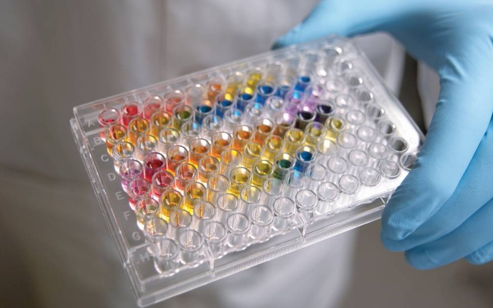
Since calcium ions are invisible to the naked eye and even to standard microscopes, scientists use ‘reporter’ molecules to make them visible. Most modern assays rely on fluorescent dyes that change their light-emitting properties when they bind to calcium. When the cell is flooded with calcium, the dye glows more brightly or changes colour, allowing a sensitive camera or plate reader to record the event. There are several types of indicators used in these processes:
- Fluorescent Indicators: These are chemical dyes like Fura-2 or Fluo-4 that are loaded into cells. They are highly sensitive and provide fast results.
- Luminescent Indicators: These use proteins like aequorin, which emit light through a chemical reaction when calcium is present, similar to how a firefly glows.
- Genetically Encoded Calcium Indicators (GECIs): These are proteins that the cell is programmed to produce itself through genetic engineering, allowing for long-term monitoring of specific tissues.
How the pharmaceutical industry uses calcium assays for safer drugs
The journey of a new drug from the lab bench to the pharmacy shelf is long and fraught with risk. One of the biggest hurdles is ensuring that a drug does not have unintended side effects on the heart. The rhythm of the heart is controlled by a precise flow of ions, and any disruption to calcium signalling can lead to dangerous arrhythmias. This is why many regulatory bodies require extensive testing of new compounds before they can move to human trials.
When researchers need to assess the functional health of cells, they frequently turn to a specialised calcium assay to gather high-resolution data on ion movement. This is especially critical when working with human induced pluripotent stem cell-derived cardiomyocytes (hiPSC-CMs). These lab-grown heart cells provide a human-relevant model for testing, and by performing an assay, scientists can see exactly how a drug candidate affects the ‘calcium transient’—the rise and fall of calcium that drives each heartbeat.
Screening for cardiac safety and ion channel activity
Beyond simple safety, these assays are used to explore the mechanisms of various diseases. For instance, in many types of heart failure, the way cells handle calcium becomes ‘leaky’ or sluggish. By using a calcium assay, researchers can test new therapies designed to fix these leaks and restore normal pumping function. The ability to monitor multiple cells simultaneously in a high-throughput format means that thousands of different chemical compounds can be screened in a single day, significantly accelerating the pace of medical discovery.
The technology behind high throughput calcium measurements
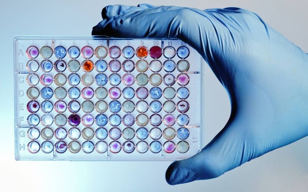
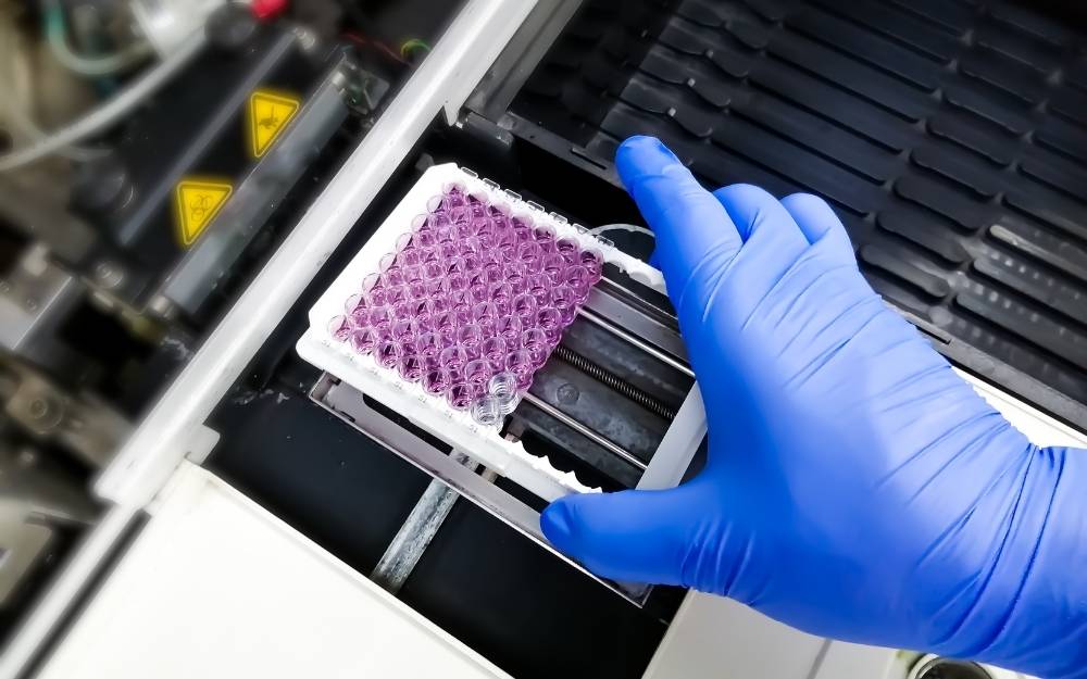
Modern laboratories do not just look at one cell at a time. To be efficient, they use high-throughput screening (HTS) platforms. These systems involve 96-well, 384-well, or even 1536-well plates, where each tiny well contains a group of cells and a different drug concentration. Specialised machines called Fluorescence Imaging Plate Readers (FLIPRs) are used to capture the data from all these wells at once.
The data produced is incredibly complex. It isn’t just a simple ‘yes’ or ‘no’ regarding calcium presence; it involves analysing the kinetics of the response. Scientists look at several factors:
- Peak Amplitude: How high does the calcium level go?
- Rise Time: How quickly does the calcium enter the cytoplasm?
- Decay Rate: How fast does the cell pump the calcium back out or into storage?
- Frequency: In beating heart cells, how many times per minute does the calcium cycle occur?
By breaking down the data in this way, researchers can identify subtle changes in cellular behaviour that might indicate a potential problem or a successful therapeutic effect. This level of detail is what makes the calcium assay an indispensable tool in modern pharmacology and cell biology.
Choosing the right indicators for your research
Not all calcium assays are created equal, and choosing the right approach depends on the specific question being asked. Some dyes are ‘ratiometric,’ meaning they are measured at two different wavelengths. This is particularly useful because it cancels out variations in dye loading or cell thickness, providing a more accurate measurement of actual calcium concentration. Other dyes are non-ratiometric and simply get brighter, which is often sufficient for high-speed screening where the primary goal is to detect a change rather than an absolute value.
Ratiometric versus non-ratiometric dyes
Ratiometric indicators like Fura-2 are the gold standard for quantitative work. They allow researchers to calculate the exact micromolar concentration of calcium within a cell. However, they require more complex optical setups. On the other hand, non-ratiometric dyes like Fluo-4 are much brighter and easier to use with standard laboratory equipment, making them the preferred choice for high-volume drug screening programmes where speed and signal-to-noise ratio are the most important factors.
Furthermore, the development of red-shifted dyes has opened up new possibilities. Because many drug compounds naturally fluoresce in the blue or green spectrum, they can interfere with traditional green dyes. Red-shifted calcium indicators allow scientists to move the measurement away from this interference, leading to cleaner data and fewer false positives in the screening process. This constant evolution in dye technology ensures that the calcium assay remains at the cutting edge of biological research, helping to organise our understanding of complex cellular networks and paving the way for the next generation of medical breakthroughs.

Vince is a medical researcher and writer with a focus on innovative therapy solutions, patient rehabilitation, and holistic treatment approaches.